Gallery
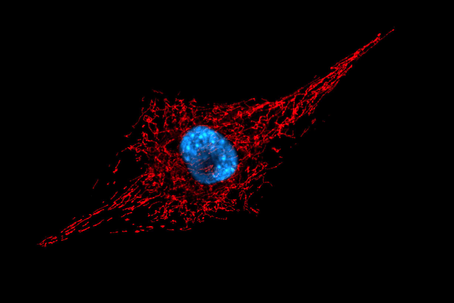
Fluorescence microscopy: Mitochondrial reticular network (stained in red with a live marker) and the nucleus (stained in blue through DNA intercalation)
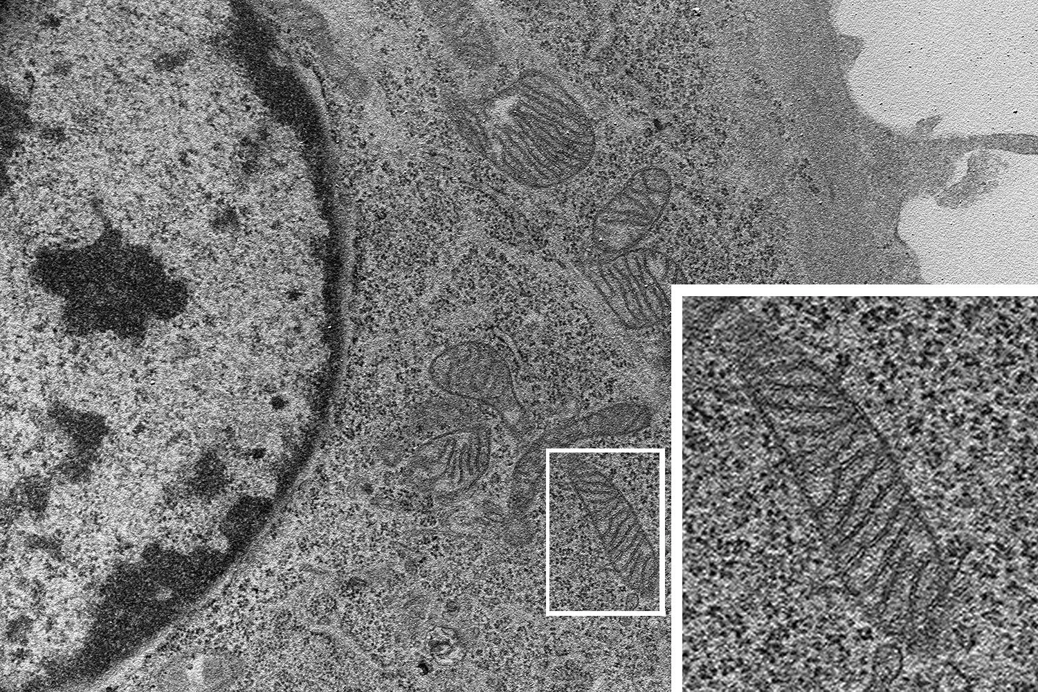
Transmission electron microscopic (TEM) image depiciting a healthy mitochondria with abundant inner membrane invaginations (cristae) and nucleus on the left.
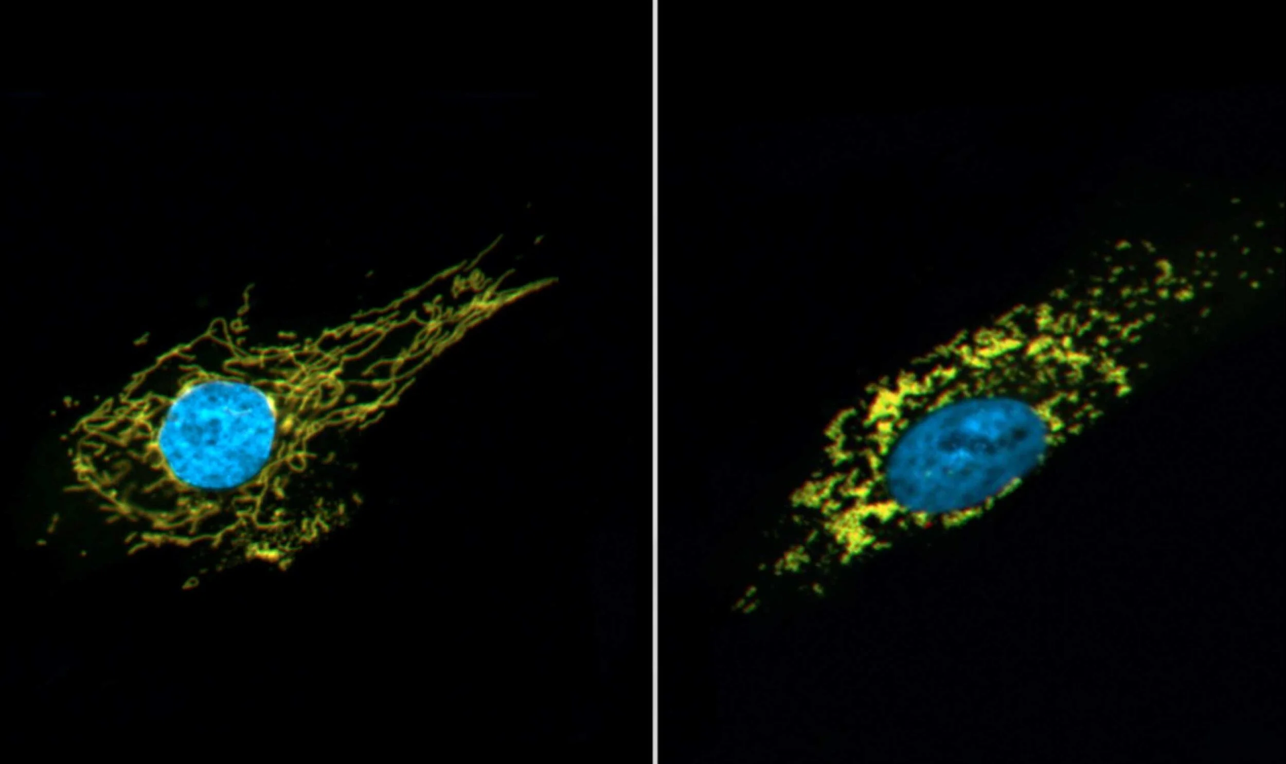
Fluorescence microscopy: Mitochondrial reticular network appearance in healthy cells visualised by a stably-expressed mitochondrial fluorescent marker in healthy (left) and stress-induced (right) states.
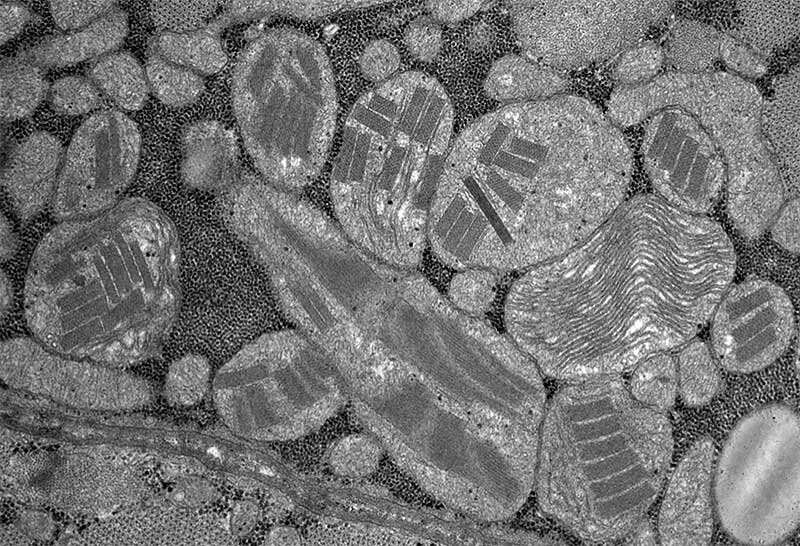
Transmission electron microscopic (TEM) image of human skeletal muscle (cross section) in a patient harbouring a mitochondrial tRNA mutation. The image shows aberrantly enlarged mitochondria with disturbed cristae and crystalline inclusions (“parking lot inclusions"). Image width approximately 10 uM.